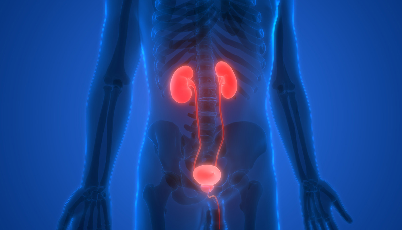
A full or partial blockage in the area where urine travels from the kidneys to the bladder is known as a ureteropelvic junction (UPJ) obstruction. This condition, more common in children than adults, affects the renal pelvis, the widened part of the ureter. Mild obstructions often resolve on their own, though a urologist may prescribe antibiotics to prevent infection.
For severe or problematic obstructions, surgery is typically recommended. The two common surgical options are endopyelotomy and laparoscopic pyeloplasty.
Diagnosis and Assessment
Patients with a ureteropelvic junction (UPJ) obstruction often exhibit symptoms such as blood in the urine (hematuria), abdominal discomfort, kidney stones, and urinary tract infections. To confirm a UPJ obstruction and exclude other potential conditions or structural issues that might hinder urine flow, various tests are conducted. These typically include blood tests, urine tests, and imaging studies to determine the severity of the obstruction.

Endopyelotomy
An endopyelotomy is a minimally invasive endoscopic procedure designed to remove the renal pelvic blockage. A special surgical balloon with an electric wire or telescope is inserted to reach the obstructed area, where scar tissue causing the blockage is excised. Due to the smaller incisions, this procedure can often be performed on an outpatient basis by a urologic surgeon.
A temporary stent is placed for four to six weeks to prevent the recurrence of scar tissue. Patients generally experience a shorter recovery time compared to traditional open surgery. During this period, antibiotics are prescribed to prevent infection. Some patients may notice blood in their urine post-surgery, which is normal and usually resolves within a few days.
Laparoscopic Pyeloplasty
For severe blockages that are unlikely to be corrected with endoscopic scar tissue removal, laparoscopic pyeloplasty may be performed. This minimally invasive procedure uses small incisions and special instruments. During the surgery, the obstruction is removed, and the affected portion of the ureter is reconnected to healthy renal pelvic tissue. The ureter is repositioned to facilitate normal urine flow.
A urology surgeon may leave a stent in place for about a week to ensure proper drainage of the ureter or use a kidney catheter (nephrostomy) instead. Alternatively, a rubber drain (Penrose drain) may be placed under the incision site. For children, a caudal or epidural nerve block is sometimes used to reduce discomfort. This procedure has a success rate of over 90 percent.
After successful surgery for UPJ, patients are monitored every three to six months with blood and urine tests and a renal ultrasound to ensure normal kidney function. Follow-up may also include specialized tests to assess urine flow. Most children treated for UPJ can resume an active and productive lifestyle once normal urine flow is restored from the affected kidney to the ureter.
Choose UCI Pelvic Health Center
Regain your health and comfort with expert ureteral and ureteropelvic junction (UPJ) treatment at UCI Pelvic Health Center. Our experienced team offers advanced, minimally invasive procedures like endopyelotomy and laparoscopic pyeloplasty to effectively address blockages and restore normal urine flow. With personalized care and state-of-the-art techniques, we ensure a quicker recovery and better outcomes.