Ureteral obstruction can be caused by a blockage either inside or outside the ureter, affecting any part of its length, including ureteropelvic junction (UPJ) obstruction or ureteral strictures at various points from the kidney to the bladder. In some cases, the obstruction is due to external pressure from a nearby mass. Risk factors for ureteral obstruction include trauma, infections, prior abdominal surgeries, radiation therapy, inflammatory conditions, and endoscopic procedures.
Ureteropelvic Junction Obstruction
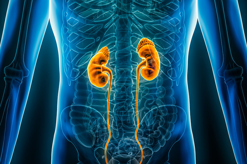
The kidneys filter salts, waste, and water from the blood, producing urine that travels through the ureters to the bladder. Proper functioning of at least one ureter is essential for urine transport.
UPJ obstruction occurs when part of the kidney is blocked, usually at the renal pelvis where the kidney connects to the ureter. This blockage can slow or stop urine flow, causing buildup and potential kidney damage. In some instances, surgery may be needed to resolve the issue.

UPJ obstruction is typically congenital, occurring in about 1 in 1,500 children and is often detected via ultrasound before birth. Symptoms in infants and children include:
- Vomiting
- Bloody urine
- Upper abdominal or back pain
- Abdominal mass
- Kidney stones
- Pain without infection
- Urinary tract infection with fever
UPJ obstruction can also develop in adults due to kidney stones, surgery complication, or upper urinary tract swelling.
Ureteral Strictures
A ureteral stricture occurs when scar tissue replaces normal tissue, narrowing the ureter lumen and reducing urinary flow, potentially leading to obstructed voiding, urinary tract infections, and urinary retention.

Diagnosing and Treating Obstructions
Diagnosis may involve blood urea nitrogen (BUN) and creatinine tests to assess kidney function. An intravenous pyelogram (IVP) can also be performed, using dye and x-rays to track urine flow. A nuclear renal scan, using radioactive material, or a CT scan in emergency situations, may also be used to diagnose the obstruction. Previous surgeries and radiation history are also assessed.
Not all cases require treatment, as some may improve over time. Necessary treatments can include open surgery or minimally invasive procedures to remove the blockage.
At the UCI Pelvic Health Center, our reconstructive urologist will create a personalized care plan based on the stricture’s location and cause. Treatment options include open surgical techniques, advanced robotic techniques, and endoscopic procedures to manage complex ureteral strictures, including those resulting from prior surgeries, urinary diversion, or radiation, as well as UPJ obstructions.
Choose UCI Pelvic Health Center
If you’re dealing with ureteral obstruction, UCI Pelvic Health Center offers expert diagnosis and personalized treatment plans. Our experienced specialists use advanced techniques to address blockages and improve urinary function.