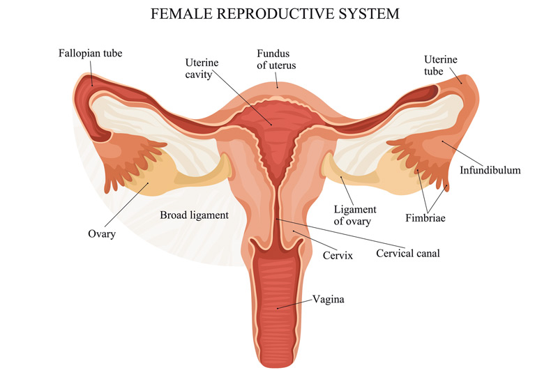
Congenital Müllerian anomalies arise when the Müllerian ducts fail to develop correctly, affecting the formation of the reproductive system, including the uterus, fallopian tubes, cervix, and the upper portion of the vagina.
Symptoms of Congenital Müllerian Anomalies
Women with Müllerian duct anomalies may experience:
- Infertility
- Pelvic pain
- Recurrent miscarriage
- Amenorrhea (absence of menstrual periods)
- Preterm labor
- Difficulty using tampons or during intercourse
Delayed diagnosis and treatment can result in chronic pelvic pain, infertility, increased retrograde menstruation, endometriosis, and missed school or work days.

Types of Müllerian Anomalies
There are around 10 types of Müllerian anomalies that affect the uterus and cervix:
1. Müllerian agenesis: Absence of the uterus, often associated with multiple reproductive abnormalities
2. Unicornuate uterus: Presence of only one half of the uterus, shaped like a banana
3. Didelphys uterus: A double uterus resulting from improper formation during fetal development
4. Bicornuate uterus: A heart-shaped uterus with an indentation at the top and one cervical opening
5. Septate uterus: A heart-shaped inner cavity of the uterus
6. Cervical agenesis: Absence of the cervix
7. Cervical duplication: Presence of two cervixes
8. Cervical hypoplasia: An unusually small cervix
9. Vaginal septum: A partition in the vagina that can be longitudinal, dividing it in half, or transverse, blocking menstrual outflow
10. Obstructed uterine horn: Formation of a uterine segment without an attached cervix or vagina
The most common anomaly is a vaginal septum, which can cause issues such as a unicornuate uterus. Symptoms depend on the septum’s location and whether the uterus has an endometrial lining. If the lining is present but the uterus is disconnected from the vagina, menstrual outflow can be blocked, causing pain. Blood accumulation in the uterine remnant or fallopian tube can also lead to pelvic masses.

Diagnosing Congenital Müllerian Anomalies
Diagnosis involves noting patient symptoms and performing a pelvic exam. Various imaging tests are used for further analysis, including:- Pelvic ultrasound
- Pelvic MRI
- Hysteroscopy: Inserting a tiny telescope through the vagina into the uterus
- Hysterosalpingography: An X-ray with dye
- Vaginoscopy: Inserting a tiny telescope into the vagina
- Laparoscopy: Inserting a thin tube with a light and camera into the abdomen